
Choosing the best Spine Surgery Microscope in 2026 is not a matter of picking the most expensive model. In a narrow surgical corridor, the surgeon needs a steady, bright view, responsive focus, and comfortable positioning. A clear image can help reveal fine anatomy. It cannot replace sound judgment, training, or careful patient selection.
Spine surgeon and microsurgical educator Dr. Richard Fessler is a relevant clinical expert to consult when evaluating these systems. However, no source article or verified quotation was provided here, so I cannot responsibly attribute a direct quote to him. That matters. A polished but invented quotation would weaken an otherwise practical comparison.
The right microscope should fit the procedure and the operating room. Consider working distance, depth of field, illumination, camera options, and how easily the optics move while the surgeon changes position. Picture a surgeon leaning over a lumbar case: a stiff arm or awkward eyepiece can become more than a small annoyance. Yet specifications do not tell the whole story. Ask how the system performs during real workflows, what training is available, and how service is handled when equipment needs attention. There is no universal winner. The best choice depends on clinical needs, team experience, budget, and support. Some comparisons will remain imperfect, because published specifications rarely capture every operating-room constraint. That is worth admitting. A careful 2026 guide should separate measurable features from marketing claims and explain where evidence remains limited.
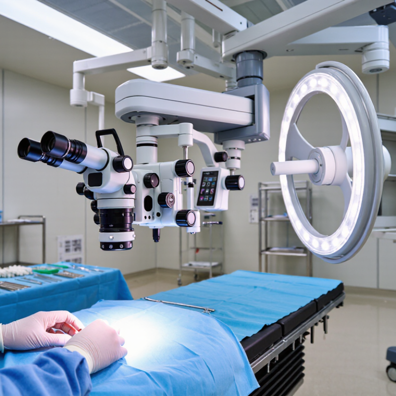
A spine surgery microscope supports precision by making small anatomical structures easier to see under controlled magnification. Clear optics and even illumination can help surgeons distinguish tissue layers around the spinal cord and nerve roots. A stable image matters: a slight movement can make a narrow working area harder to interpret. The microscope does not make decisions, but it can give the surgical team a more detailed view.
Useful features include adjustable magnification, focused lighting, and smooth positioning that lets the surgeon change the viewing angle without losing the target. Depth perception is important when working through a small incision or along a deep surgical corridor. Controls should be reachable without forcing an awkward posture. Small shifts count. A well-positioned eyepiece and balanced equipment can reduce strain during long procedures, though comfort depends on the room, the surgeon, and the case.
The best choice in 2026 is not necessarily the system with the most features. It should fit the procedure, operating-room layout, and team’s training. Staff need time to practice focusing, repositioning, and keeping the field clear. Even good equipment can feel awkward at first. That deserves attention, not dismissal: setup and familiarity affect how consistently a microscope supports precise work.
The best microscope is the one that supports consistent visualization in your actual operating room. Compare image clarity, depth of field, illumination, and working distance using a spine model or representative procedure setup. Check whether the microscope stays stable when repositioned and whether controls are easy to reach while wearing gloves. Small details matter.
Ergonomics deserve equal attention. Surgeons should be able to maintain a neutral neck and shoulder position, while assistants retain a clear view. Evaluate camera output, recording options, and compatibility with existing room equipment. The WHO’s 2024 Global Patient Safety Report estimates that one in ten patients experiences harm in healthcare; this broad figure is not specific to microscopes, but it underscores the value of dependable workflows. A microscope cannot guarantee safer surgery. Nor can a showroom demonstration reveal every limitation.
Spine surgery microscope systems differ less by headline magnification than by how clearly they support the surgeon’s view during a specific procedure. Traditional optical systems provide direct stereoscopic vision and familiar controls. Digital systems display a magnified view on a monitor, which can help the whole team follow the field. Hybrid designs combine these approaches, though added features may also mean more setup and training.
Compare image depth, illumination, working distance, and how easily the microscope moves between positions. In a narrow operative field, stable depth perception and even lighting can make small structures easier to distinguish. Check whether the arm can be repositioned without disrupting the surgeon’s posture. A monitor may improve shared viewing, but it does not automatically replace the value of direct binocular vision. That depends on the operator and the case.
Ergonomics deserve close attention. Long procedures can make awkward viewing angles costly, even when the optics are excellent. Test controls with gloves, assess whether assistants can see the display, and ask how routine cleaning and service affect availability. Training matters, too. A sophisticated system is not necessarily the best fit if the team cannot use it confidently. There is no universal winner; hospital workflow, surgeon preference, procedure mix, and budget all shape the comparison. Some priorities may conflict. That part is easy to underestimate.
There is no single best system for every spine procedure. This comparison describes common system types rather than specific products; features and performance vary by configuration. Confirm capabilities with the manufacturer and assess them in a hands-on demonstration.
| System type | Visualization and depth perception | Ergonomics and team view | Potential advantages in spine surgery | Trade-offs and checks before purchase | Potential best fit |
|---|---|---|---|---|---|
| Conventional optical surgical microscope | Direct binocular viewing through eyepieces; optical magnification and illumination support detailed work in a small operative field. | The surgeon generally looks through oculars. Assistant viewing depends on the configuration and may require an assistant scope or other accessories. | Familiar workflow and direct optical viewing can suit microsurgical tasks such as nerve-root and dural work. | Check working distance, field of view, illumination, focus controls, movement range, and whether the viewing posture is comfortable for the intended approach. | Teams that prefer eyepiece-based viewing and need a microscope for established microsurgical workflows. |
| Motorized optical microscope | Uses optical binocular viewing; motorized movement or focus functions may be available, depending on the configuration. | Motorized positioning can reduce some manual adjustments. Ergonomic benefit depends on controls, balance, reach, and how the microscope is set up. | Can support frequent repositioning across a surgical field, including during procedures that require changing viewing angles. | Verify which movements are motorized, how controls are operated, the time needed to reposition, and compatibility with the operating-room layout. | Surgeons who value optical viewing but want powered positioning or focus features. |
| Digital 3D exoscope | Displays a magnified surgical view on a monitor; 3D depth perception is available in systems designed for stereoscopic viewing and requires compatible viewing equipment. | The surgeon and team can view the same monitor image. A screen-based workflow may allow a more upright head and neck posture, depending on monitor placement. | Shared visualization can make it easier for the surgical team to follow the field; may suit teaching and procedures where screen viewing is preferred. | Assess image latency, monitor position, depth perception, focus and zoom controls, lighting, and performance at the working distance required for spine procedures. | Teams seeking shared screen-based visualization and a potential alternative to prolonged eyepiece viewing. |
| Hybrid optical and digital system | Combines optical viewing with a digital display or recording pathway, where included in the configuration. | May offer eyepiece viewing for the primary surgeon and a monitor view for other team members; available options vary. | Can accommodate mixed preferences for direct viewing and shared display, without requiring every team member to use the same viewing method. | Clarify whether the digital image is simultaneous, what accessories are required, and whether switching between viewing modes interrupts workflow. | Departments with surgeons or procedures that benefit from both eyepiece and monitor-based viewing. |
| Image-guided microscope integration | May combine microscope visualization with navigation or imaging information displayed in the field or on a separate screen; integration depends on compatible equipment. | Can bring relevant visual information into the team’s workflow, but may add setup steps and require coordination among systems. | Potentially useful when the procedure already uses navigation or other image-guidance tools; it does not replace clinical judgment or standard verification steps. | Confirm supported interfaces, registration workflow, display location, setup time, staff training needs, and what happens if integration is unavailable. | Spine teams that routinely use compatible navigation or imaging systems and can support the added workflow. |
Choosing the best spine surgery microscope in 2026 starts with the procedure, not the feature list. The WHO’s 2023 low-back-pain fact sheet reports 619 million people affected in 2020. A 2023 Global Burden of Disease analysis projects 843 million by 2050. These figures describe disease burden, not which microscope a surgeon needs.
For lumbar microdiscectomy, prioritize coaxial illumination, stable fine focus, and enough working distance to keep instruments clear in a deep, narrow corridor. Smooth magnification changes help when moving between nerve-root detail and the wider field. Fit matters.
For decompression or instrumented fusion, a broader field, rapid zoom, assistant viewing, and clear camera output may support team communication and documentation. Check that the microscope arm stays steady when repositioned, and test controls from the surgeon’s actual operating posture.
A specification sheet cannot reproduce a crowded operating room. A feature can look impressive and still slow the case. Compare the view at the working distance used for the planned procedure, then assess lighting, depth perception, drape clearance, and compatibility with the team’s workflow.
Not always. Microscope choice also depends on surgeon training and institutional setup; published population estimates cannot establish one universally superior model. A practical evaluation should include surgeons and operating-room staff, with procedure-specific trials rather than a single showroom demonstration.
What Is the Best Spine Surgery Microscope in 2026?
Practical Considerations for Choosing and Implementing a System
The right microscope is the one that supports the team’s actual procedures and operating-room layout. During evaluation, test image clarity at the working distance used for spinal exposure. Check depth of field, illumination, and how quickly the view can be repositioned. Small delays matter. Ask surgeons and staff to assess the controls while wearing gloves, not just during a showroom demonstration.
Fit the system to the room. Measure ceiling clearance, table movement, and space around carts before selecting a configuration. Confirm that assistants can see the field and that the microscope does not obstruct access to monitors or instruments. A useful trial includes a realistic setup, routine draping, and a simulated change in patient position. It may reveal awkward steps that a specification sheet misses.
Implementation needs planning, too. Arrange hands-on training for surgeons, scrub staff, and technicians, then establish clear cleaning and maintenance routines. Check service response times, replacement-part availability, and the full cost of ownership. Record setup time and staff feedback during early cases. A trial can still be misleading if it uses an unusually spacious room; that is an easy detail to overlook. The final choice should reflect local workflow, not just impressive optics.
Practical Considerations for Choosing and Implementing a System
How to use this: With the optical configuration held constant, field diameter is approximately inversely proportional to magnification. The values shown are normalized optical calculations, not specifications for a particular microscope. When comparing systems, also assess working distance, illumination, image quality, ergonomics, assistant viewing, and ease of integration into the operating room.
