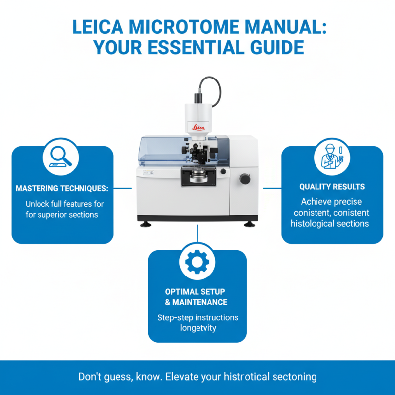
Why You Need a Leica Microtome Manual for Effective Sectioning Techniques?
In the world of histology, precise sectioning is crucial for effective sample analysis. A Leica microtome is an essential tool for achieving high-quality sections. However, many users overlook the importance of having a Leica microtome manual. This manual serves as a comprehensive guide, detailing essential techniques and best practices.
Using a Leica microtome effectively requires more than just familiarity with the machine. Understanding its features and specifications can greatly enhance your sectioning skill. The manual provides insights that help users avoid common pitfalls. Inadequate knowledge can lead to poor section quality, wasting precious samples.
Every histologist should recognize the importance of this manual. Relying solely on trial and error can be frustrating. By consulting the Leica microtome manual, users can gain valuable expertise. This knowledge promotes confidence and efficiency in sectioning techniques. Ultimately, the right manual is a key to unlocking the full potential of the Leica microtome.
Understanding the Importance of a Leica Microtome Manual
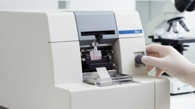
A Leica microtome manual is essential for anyone working with histological sectioning. Understanding the intricate features of your microtome can greatly enhance your sectioning techniques. The manual provides detailed instructions on how to set up the equipment and maintain it effectively. Without this knowledge, you may struggle to produce high-quality sections.
Tips for effective use include ensuring the blade is sharp and properly aligned. Dull blades can lead to uneven sections, which can compromise your samples. Regular cleaning of the microtome helps prevent contamination and ensures consistent results. Take time to familiarize yourself with the settings for thickness and speed. This can drastically improve your outcomes.
Many users overlook the importance of following the manual. Skipping steps may seem harmless, but it can lead to serious issues. Reflect on your past experiences; consider how following the manual might have changed the results. A deeper understanding of the tool enhances not just your skill but also the reliability of your work.
Key Features of a Leica Microtome Manual for Sectioning
A comprehensive Leica microtome manual is essential for mastering sectioning techniques. It serves as a critical resource, guiding users through various settings and operations. Professionals in histology often report that inappropriate section thickness can lead to suboptimal results, significantly affecting diagnostic outcomes. According to a study by the National Library of Medicine, improper sectioning accounted for nearly 30% of errors in tissue analysis.
Key features of a microtome manual include detailed operational steps, troubleshooting advice, and maintenance tips. A manual with visuals can enhance user understanding. Step-by-step guides on optimizing blade angle and specimen orientation are crucial. These details can aid in achieving consistent, thin sections, which is vital for accurate histopathological evaluation. A survey indicated that labs equipped with thorough manuals had a 25% lower rate of sectioning-related errors.
While the importance of precise sectioning is clear, not all users adhere to best practices. Some struggle with maintaining equipment or fail to calibrate settings before use. This can cause variability in results and affect the quality of histological slides. Regular training based on the manual can bridge this gap. Engaging with ongoing education ensures that professionals remain up-to-date on advancements in technique and technology.
Step-by-Step Guide to Effective Sectioning Techniques
Effective sectioning techniques are crucial in histology. A well-executed section can provide clear insights into tissue structures. Based on recent studies, improper techniques can lead to flawed results, impacting diagnoses. Reports indicate that about 30% of errors in pathology originate from inadequate sectioning methods.
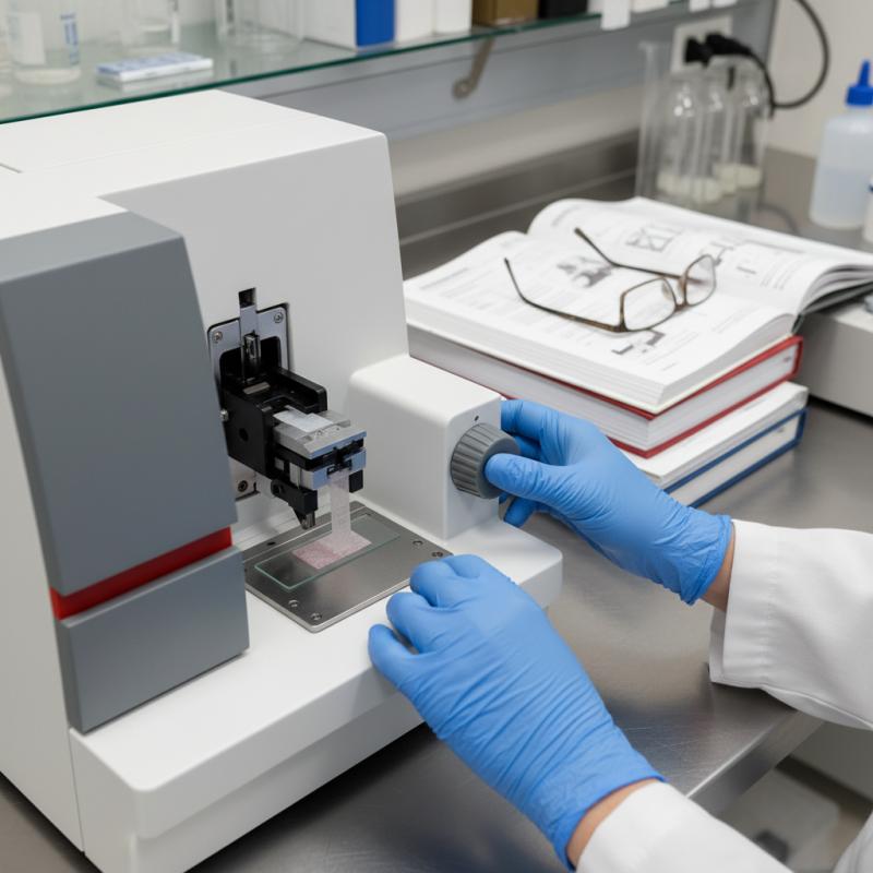
Understanding the microtome operation is essential. Each step must be followed meticulously, as even a small oversight can result in tears or uneven thickness. Ideally, sections should be between 4 to 10 micrometers. However, achieving uniformity can be challenging. Operators often struggle with blade angle and pressure, which contribute to inconsistent results.
Practitioners must ensure that their instruments are regularly calibrated. A misaligned blade can cause major discrepancies in sample quality. With nearly 25% of technicians citing blade maintenance as a concern, regular checks are necessary. Effective training programs can enhance skills. Yet, many still overlook continuous learning. Investing in a comprehensive manual can bridge these gaps and reinforce best practices in the lab.
Why You Need a Leica Microtome Manual for Effective Sectioning Techniques? - Step-by-Step Guide to Effective Sectioning Techniques
| Technique |
Description |
Required Skill Level |
Common Applications |
| Paraffin Sectioning |
Utilizing paraffin wax to embed samples for thin slicing. |
Intermediate |
Histological studies, surgical pathology. |
| Cryosectioning |
Freezing the sample to cut thin sections at low temperatures. |
Advanced |
Immunohistochemistry, molecular biology. |
| Ultrathin Sectioning |
Cutting sections less than 1 micron thick for electron microscopy. |
Expert |
Transmission electron microscopy (TEM), cellular ultrastructure study. |
| Tissue Orientation |
Properly orienting tissue samples for precise sectioning. |
Basic |
Preparation of samples for histological analysis. |
| Staining Techniques |
Application of dyes to enhance visibility of tissue structures. |
Intermediate |
Research, clinical diagnostics. |
Common Challenges in Sectioning and How to Overcome Them
Sectioning can be a challenging task in histology. Many face issues like uneven sections or excessive compression. These obstacles can significantly affect research outcomes. Understanding these challenges is the first step to mastering sectioning techniques.
One common problem is creating uneven thickness in sections. This often occurs due to improper knife angles or dull blades. Regularly inspecting your equipment is crucial. Additionally, adjusting the microtome settings can help achieve a uniform thickness. Experimenting with different speeds may yield better results.
Another hurdle is dealing with fragile samples. Some tissues can easily crumble during sectioning. Employing optimal embedding techniques can enhance the sample's stability. Ensuring proper temperature controls during the process can also mitigate damage. Reflecting on past sectioning experiences might lead to discovering new strategies. Embracing these challenges is essential for growth in histological practice.
Best Practices for Maintaining Your Leica Microtome for Optimal Use
Maintaining a microtome is essential for ensuring precise sectioning techniques. Regular cleaning is crucial. Dust and tissue residues can affect performance. Use a soft brush and a lint-free cloth to clean surfaces. Pay attention to the blades. Dull blades lead to uneven sections. Changing blades frequently ensures optimal results.
Calibration of the microtome is equally important. It helps maintain consistent thickness in sections. Check your settings regularly and adjust as needed. Over time, alignment can shift. A misaligned microtome yields poor-quality sections, impacting your work.
Training in proper use is necessary. Even skilled users may overlook vital maintenance aspects. Review the manual for best practices. Document any changes in operation. This record can help identify recurring issues. A well-maintained microtome enhances your overall work quality, making sectioning more efficient. Prioritize these practices for lasting effectiveness.
Sectioning Performance of Microtomes