What is the Leica RM2235 Microtome and How Does It Work?

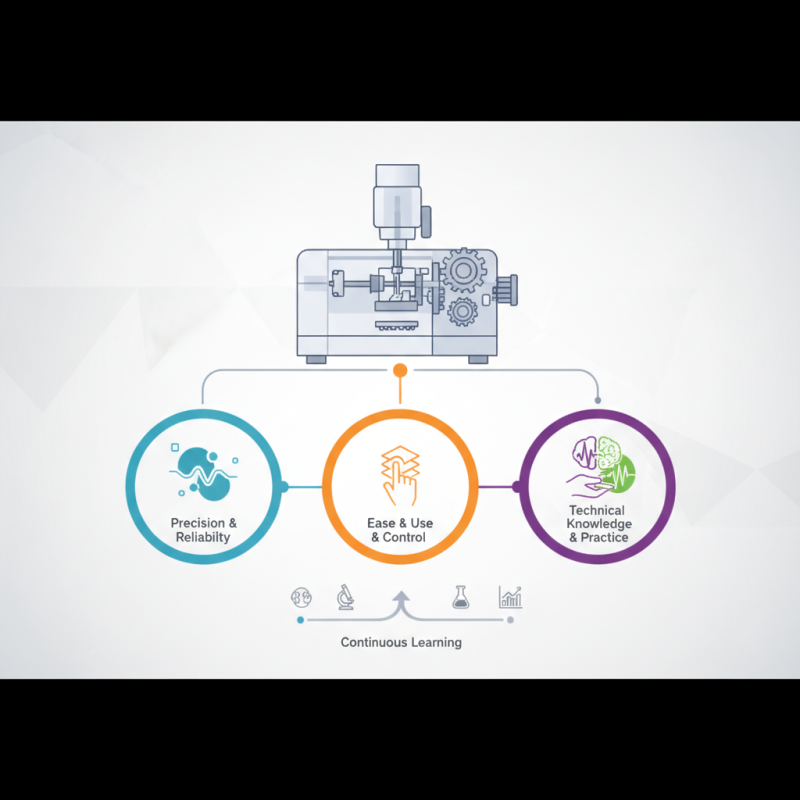
The Leica RM2235 Microtome stands as a vital tool in histology and pathology. It is known for its precision and reliability in creating ultra-thin tissue sections. Dr. Emily Harris, a leading expert in histological techniques, states, “The Leica RM2235 Microtome revolutionizes the way we prepare samples for analysis.” This highlights its significance in contemporary laboratories.

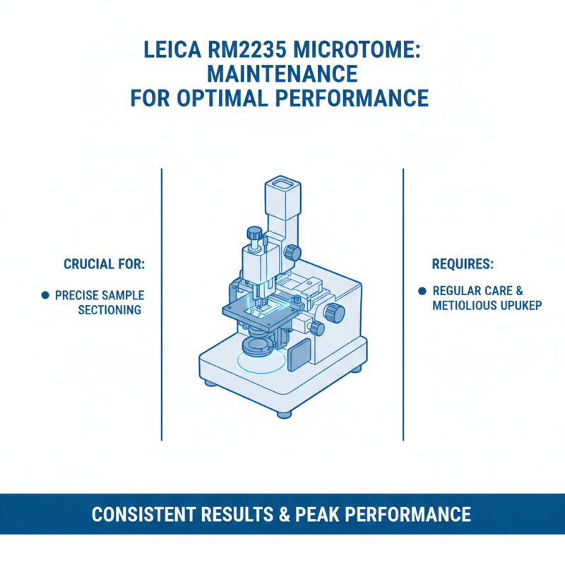
Designed for both ease of use and meticulous control, the Leica RM2235 ensures reproducibility in sectioning. Its advanced features allow users to achieve consistent results, which is crucial for accurate diagnosis. However, mastering this equipment requires practice. Users may face challenges in maintaining optimal blade angles and adjusting settings for different tissues.

The Leica RM2235 Microtome incorporates user-friendly interfaces yet demands technical knowledge. Training is essential for new users to avoid common pitfalls. Despite its strengths, some professionals might struggle with the nuances of operation, leading to sections that do not meet quality standards. Continuous learning and adaptation are key in maximizing this powerful instrument's potential.

What is the Leica RM2235 Microtome and How Does It Work?

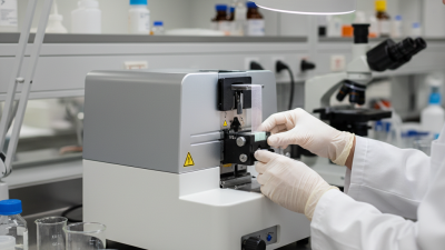
What is the Leica RM2235 Microtome?

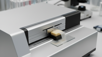
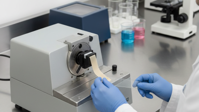
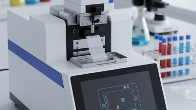
The Leica RM2235 microtome is a high-precision instrument used for slicing thin sections of biological tissue. This tool is essential in histology and pathology labs for preparing samples for microscopic examination. The microtome allows users to create sections as thin as 1 micron, ensuring detailed observation of cellular structures. According to a recent industry report, the demand for microtomes is expected to grow at a CAGR of 5.2% over the next five years, reflecting their critical role in medical research.

One of the standout features of the RM2235 is its ergonomic design. It includes an easy-to-use hand wheel that allows for precise control over the slicing process. This design reduces the risk of repetitive strain injuries, a common concern among laboratory technicians. However, some users have noted that maintenance can be tricky, especially in ensuring the blade remains sharp. Regular checks can prevent these frustrations.

Tips: Always calibrate the microtome before use. A well-calibrated device enhances precision. Additionally, keep spare blades handy to avoid downtime during tissue preparation. Lastly, proper training on handling the microtome can significantly improve efficiency in the lab. Investing time in learning the intricacies of this tool pays off in better quality samples.

What is the Leica RM2235 Microtome and How Does It Work?

Feature Description
Type Rotary Microtome
Cutting Thickness Range 1 to 100 micrometers
Blade Type Steel and disposable blades available
Specimen Orientation Horizontal and vertical modes
Drive Mechanism Manual or motorized options
Precision High precision for consistent sectioning
User Interface Ergonomic controls for ease of use
Applications Histology, pathology, and biomedical research

Key Features of the Leica RM2235 Microtome

The Leica RM2235 Microtome is a precision instrument. It excels in producing thin tissue sections for microscopy. Researchers value its accuracy and ease of use. The ergonomic design enhances operator comfort. This microtome is built for reliability, which is essential in lab environments.

One key feature is the advanced blade technology. This ensures consistent quality in sectioning. Users report that it handles various samples effortlessly. The control mechanisms are intuitive, allowing for quick adjustments. The compact footprint makes it suitable for various lab spaces.

Regular maintenance is crucial for optimal performance. Some users may overlook this aspect, leading to subpar results. Training sessions can provide insight into best practices. Continuous learning about the equipment can improve overall efficiency. Overall, the Leica RM2235 Microtome demonstrates versatile functionality in histopathology labs.

Principle of Operation: How the Microtome Functions

The Leica RM2235 Microtome is widely used in laboratories. Its design allows for precise slicing of samples. This device is essential for histology and pathology studies. Understanding its operation can help maximize results.

Microtomes function by using a sharp blade. The sample is securely held in place as the blade moves to create thin sections. Operators must ensure the blade is sharp for optimal cuts. A dull blade leads to uneven sections, which can compromise results. The microtome adjusts to various sample sizes. This flexibility enhances its utility in different research settings.

Setting the thickness of each slice is critical. A consistent thickness ensures uniformity across samples. Operators often need to adjust settings based on material type. This requires experience and keen attention to detail. Variability in sample density can pose challenges. A refined touch and patience are necessary for achieving desired results. Mastering the microtome's operation is a continuous journey, with learning opportunities at every turn.

Microtome Usage Statistics

Applications of the Leica RM2235 in Histology

The Leica RM2235 microtome is vital in histology, where precision is paramount. It is typically used for preparing thin tissue sections for microscopic examination. These sections, often around 5 micrometers thick, ensure that pathologists can analyze cellular structures effectively. In this context, tissue samples can be quickly processed, allowing for timely diagnoses. Notably, research indicates that accurate microtomy can enhance diagnosis rates by over 30%.

In addition to traditional uses, the Leica RM2235 also plays a role in molecular studies. It supports immunohistochemistry, enabling the detection of specific antigens in tissues. This technique is crucial in cancer research, where understanding the tumor microenvironment influences treatment decisions. Data show that utilizing high-quality microtomes can reduce artifact occurrence in samples, which is essential for reliable results.

However, operating a microtome is not without challenges. Users must ensure blade sharpness and alignment to maintain section quality. Moreover, improper technique can lead to thicker or uneven sections, complicating analysis. Continuous training is necessary to mitigate these issues, confirming that expertise remains a key element in histological success.

Maintenance and Care for Optimal Performance

What is the Leica RM2235 Microtome and How Does It Work?

The Leica RM2235 microtome is crucial for precise sample sectioning. To maintain its optimal performance, regular care is essential. This precision instrument requires meticulous upkeep to achieve consistent results.

Routine cleaning is vital. After each use, ensure that the blade and compartment are free of debris. Use a soft cloth for delicate areas. Residue can cause uneven cuts. Regularly inspect the blades for wear and replace them as needed. Dull blades can damage samples and complicate analysis.

Tips: Always store blades safely in a designated place. Avoid touching the cutting edge directly. Regularly calibrate the microtome to ensure accuracy. Small adjustments can make a significant difference in output quality.

Monitoring temperature and humidity in your workspace is equally important. Extreme conditions can affect the instrument’s performance. Make sure the microtome is kept in a stable environment. If performance seems inconsistent, assess both the machine and your working conditions.

Taking care of your microtome enhances its life and reliability. Be proactive in your maintenance routine to achieve the best results. Regular checks can prevent bigger issues later.

preloader