What Is an HM 325 Microtome and How Does It Work?

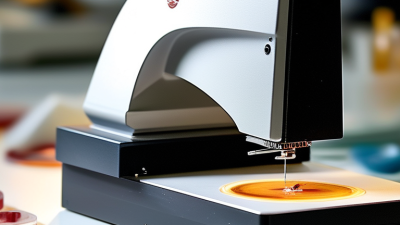
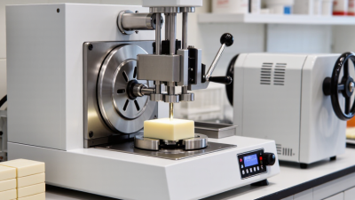
The hm 325 microtome is a rotary instrument used to cut extremely thin tissue sections. It prepares paraffin-embedded specimens for microscopic examination. Its purpose is simple. Its accuracy is not.

According to the World Health Organization’s GLOBOCAN 2022 estimates, nearly 20 million new cancer cases occurred worldwide. About 9.7 million people died from cancer that year. These figures show why dependable histology remains essential. Before a pathologist can assess cellular structure, a specimen must be processed, embedded, sectioned, stained, and examined. The hm 325 microtome supports the sectioning stage.

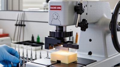
Its operation depends on controlled mechanical movement. The operator secures a tissue block in the specimen holder. A handwheel advances the block toward a sharp disposable blade or fixed knife. Each rotation produces a measured slice, often only a few micrometres thick. The section then floats on a warm water bath before moving onto a glass slide. Small errors matter.

A loose clamp may create chatter. Excessive pressure can compress the tissue. A damaged blade may produce uneven ribbons. The College of American Pathologists emphasizes equipment control, specimen quality, and reproducible technical procedures through its laboratory accreditation checklists. These expectations place the instrument within a wider quality system, not as a standalone solution. In real laboratories, experience still matters. The hm 325 microtome can provide reliable sections, but only when maintenance, blade selection, block temperature, and operator technique work together. Its design appears straightforward. The results can still demand patience, judgment, and occasional correction.
What Is an HM 325 Microtome and How Does It Work?

HM 325 Design: Rotary Microtome Components and 64 mm Vertical Stroke

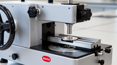
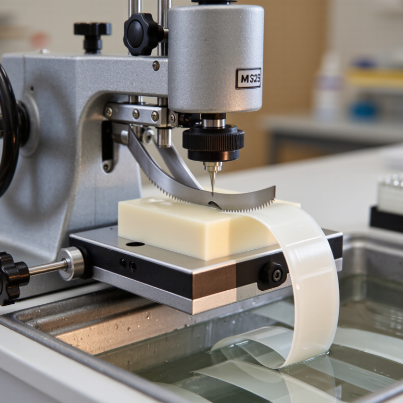
An HM 325 microtome is a rotary instrument for cutting thin tissue sections. Its design supports controlled movement around a fixed cutting position. The main components include a handwheel, specimen clamp, feed mechanism, knife holder, coarse advance, and safety lock. Together, they convert circular handwheel motion into precise vertical travel.

The 64 mm vertical stroke is the key design detail. It describes the specimen’s up-and-down movement during one cutting cycle. As the handwheel turns, the tissue block rises toward the blade, passes across its edge, then returns below it. The feed mechanism advances the block by the selected thickness. This repeated motion creates ribbons for mounting on glass slides. The stroke is substantial, but it is not the section thickness.

IARC’s Global Cancer Observatory recorded about 20 million new cancer cases worldwide in 2022. It projects annual cases could reach 35 million by 2050. That workload increases pressure on reliable sample preparation. In practice, stable clamping and smooth feed control matter more than impressive specifications alone. A loose cassette can produce chatter, compression, or uneven ribbons. The 64 mm movement may look generous, yet perfect sections still depend on blade condition, paraffin quality, and operator technique. I would not treat the specification as a guarantee. It is only one part of the result.

Specimen Clamping: Positioning Blocks up to 50 × 50 mm

What Is an HM 325 Microtome and How Does It Work?

An HM 325 microtome is a precision instrument for cutting thin sections from embedded specimens. It advances a clamped block toward a sharp blade. Each controlled stroke removes a narrow section, often for microscopic examination. The specimen clamping system accepts blocks up to 50 × 50 mm, giving technicians useful flexibility with larger samples.

Correct positioning matters more than simple size compatibility. The block should sit firmly in the holder, with its cutting face parallel to the blade edge. Uneven clamping can cause chatter, compression, or sections that vary in thickness. Before cutting, inspect the block surface and remove loose wax. A small tilt can become obvious after several strokes. Even experienced users recheck alignment; that extra moment often prevents wasted sections.

Tips: Tighten the clamp securely, but avoid crushing fragile embedding material. Center smaller blocks whenever possible. Use steady hand movement and adjust thickness gradually. If ribbons break, check blade condition, room temperature, and block alignment before changing several settings at once. Record what worked. In practice, one adjustment at a time is easier to evaluate, although busy laboratories sometimes overlook this simple discipline.

Section Setting: Selecting 0.5–60 µm Thickness for Histology

What Is an HM 325 Microtome and How Does It Work?

An HM 325 microtome is a rotary instrument for cutting thin tissue sections. Its section setting spans approximately 0.5–60 µm. This range supports different histology tasks, from delicate sections to thicker trimming cuts. Paraffin tissue commonly works between 2 and 7 µm. A 3–5 µm setting often produces clear cellular detail for routine light microscopy. Thicker sections may help when tissue is fragile or poorly supported.

The selected thickness controls how much material reaches the slide. At 0.5–1 µm, even small setting errors can affect section continuity. These very thin cuts also require firm embedding and a sharp, properly positioned blade. For initial trimming, 10–30 µm can remove excess paraffin efficiently. A 60 µm setting is useful for rapid block reduction, not usually for final diagnostic sections. The handwheel advances the specimen toward the blade by the selected distance. Each rotation creates a controlled slice.

In practice, thickness is only one part of section quality. Block temperature, wax hardness, blade clearance, and cutting speed also matter. I would begin with a moderate setting, then adjust after examining the ribbon. The first setting is rarely perfect. Excessive compression may suggest a dull blade or warm block. Chattering can indicate vibration, weak clamping, or unsuitable clearance. A clean ribbon, even thickness, and visible nuclei provide better evidence than the dial alone. Small adjustments often outperform dramatic changes.

Cutting Process: Converting Handwheel Rotation into Uniform Sections

What Is an HM 325 Microtome and How Does It Work?

An HM 325 microtome produces thin, repeatable sections for microscopic examination. Its cutting process begins with the handwheel. Each rotation moves the specimen holder through a controlled vertical path beside the knife. At the same time, an internal feed mechanism advances the specimen by a selected thickness. This coordination turns manual movement into measured sectioning. Small adjustments matter.

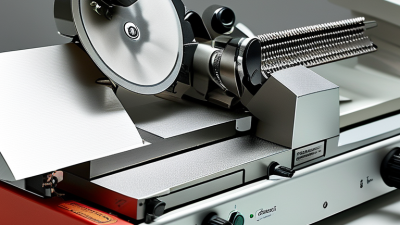
During the cutting stroke, the block meets the knife edge at a stable clearance angle. The blade removes a narrow layer instead of tearing the surface. On the return stroke, the specimen moves away from the blade, protecting the new face. A ratchet or lead-screw system controls the next advance. The operator should turn the handwheel smoothly, without sudden stops. Uneven force can create compressed, curved, or incomplete sections. The process looks mechanical, yet judgment remains important. I still recheck the feed setting when sections change unexpectedly. That extra check often reveals a loose clamp or unsuitable block hardness.

Tips: Secure the specimen firmly before cutting. Keep the knife edge clean and inspect it under good lighting. Begin with a slightly thicker setting, then reduce it gradually. Trim the block face evenly before collecting sections. If ribbons become irregular, pause and inspect the blade, clearance angle, and handwheel rhythm. Do not assume the machine is at fault. Sometimes the operator’s movement is the variable.

Rotary Microtome Cutting Process: From Handwheel Rotation to Uniform Sections

The chart shows the theoretical number of sections produced from a 100 µm block at different nominal section-thickness settings.

A rotary microtome converts controlled handwheel movement into repeated specimen advances toward a fixed blade. At a 1 µm setting, a 100 µm block can theoretically produce 100 sections; at thicker settings, fewer sections are obtained. Actual yields may be lower because of trimming, tissue loss, compression, and section defects.

Quality Control: Managing 1–30 µm Sections for Routine Microscopy

What Is an HM 325 Microtome and How Does It Work?

An HM 325 microtome cuts embedded tissue into controlled sections for routine microscopy. Its handwheel advances the specimen block, while a sharp blade removes a thin ribbon. The practical range is 1–30 µm. Routine paraffin work usually stays near 3–5 µm, according to established histotechnology guidance. Thicker sections can support connective-tissue studies, while thinner sections improve nuclear detail. Small changes matter. A 1 µm error may alter cell density and staining interpretation.

Quality control begins before cutting. ISO 15189:2022 requires laboratories to monitor equipment performance, document maintenance, and investigate nonconforming results. The College of American Pathologists’ 2024 accreditation checklist also emphasizes traceability, section quality, and corrective action. Record section thickness, block temperature, blade condition, and operator adjustments. Check ribbons under reflected light. Compression, chatter, knife lines, and uneven thickness are visible warnings. I still treat the first ribbon as evidence, not proof. Tissue hardness can change across one block.

Tips: Let the block equilibrate before trimming. Use consistent handwheel speed. Replace damaged blades early. Measure questionable sections with a calibrated method. Keep a control log with date, thickness, artifact, and corrective action. A clean-looking section can still be too thick. That is an easy mistake to miss.

What Is an HM 325 Microtome and How Does It Work? — Quality Control: Managing 1–30 µm Sections for Routine Microscopy

Section Thickness Typical Application Operating Principle Recommended Quality Criteria Common Section Defects Corrective Action
1–2 µm Very thin resin sections and selected high-resolution light-microscopy work. The specimen advances toward a stationary blade by a precisely controlled feed mechanism; each handwheel rotation produces a uniform cut. Specialized
Continuous ribbons or intact sections; minimal compression; no chatter lines.
Compression, knife marks, tearing, and incomplete section transfer. Use a sharp, suitable blade; stabilize the block; reduce cutting speed; verify clearance angle and specimen hardness.
3–5 µm Thin paraffin sections for detailed tissue morphology and special stains. The rotary mechanism repeatedly moves the specimen through a fixed cutting plane while the selected feed determines section thickness. Preferred
Even thickness, complete tissue representation, clean ribbons, and minimal folds.
Wrinkles, folds, knife lines, and variable thickness. Adjust water-bath temperature, replace or reposition the blade, cool the block if needed, and confirm that the block face is correctly trimmed.
6–8 µm Routine paraffin histology and general tissue morphology. Repeated forward movement of the specimen and return movement of the handwheel create sequential sections at the selected thickness. Preferred
Sections should float flat, retain tissue architecture, and show consistent thickness across the block.
Thick-and-thin areas, chatter, holes, and ribbon separation. Check block orientation and clamp security; reduce vibration; ensure adequate paraffin infiltration and a clean blade edge.
9–12 µm Routine sections from harder, dense, or partially decalcified paraffin blocks. A larger feed setting produces thicker sections, which can improve handling of firm or uneven specimens. Conditional
Sections must remain intact and sufficiently transparent for the planned stain and microscopy method.
Compression, scoring, tearing, and excessive background thickness. Soften or cool the block as appropriate; reduce cutting speed; inspect decalcification quality; use a blade suited to the specimen.
13–20 µm Thicker sections for selected connective tissue, whole-mount, or demonstration preparations. The feed mechanism advances the specimen farther for every cutting cycle, producing a thicker section that requires careful flattening. Conditional
Section integrity and correct specimen orientation are more important than maximum optical resolution.
Uneven flattening, folds, tearing, and poor stain penetration. Use a clean flotation bath, avoid overheating, allow adequate deparaffinization, and verify that the selected thickness suits the staining protocol.
21–30 µm Thick sections for selected demonstration, serial-section, or three-dimensional morphology workflows. The increased feed produces a robust section, but the thicker layer may require slower handling and longer reagent access during staining. Specialized
No major tears or folds; staining should be sufficiently even for the intended observation method.
Incomplete flattening, trapped water, patchy staining, and section detachment. Use adhesive-coated slides when appropriate; extend reagent times based on validation; handle sections gently and confirm compatibility with the imaging method.

Quality-control checks: Before routine cutting, confirm secure specimen clamping, correct block orientation, a clean and undamaged blade, stable instrument movement, and a verified thickness setting. Record section thickness, operator, specimen type, blade condition, cutting artifacts, and any corrective action.

preloader