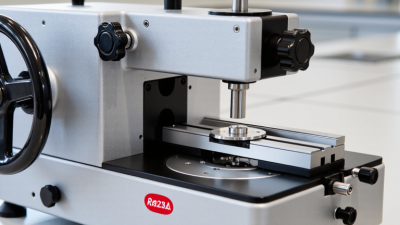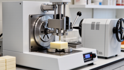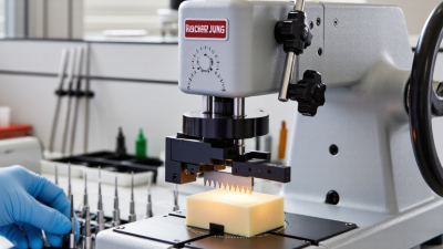
The Leica RM2235 microtome is a manual rotary instrument used to cut thin tissue sections for histological examination. It typically trims paraffin-embedded blocks into ribbons, often between 2 and 5 micrometres thick. These sections can be floated on a warm water bath, mounted on glass slides, stained, and examined under a microscope.
Its practical value becomes clearer in diagnostic pathology. The International Agency for Research on Cancer estimated approximately 20 million new cancer cases worldwide in 2022, increasing the need for reliable tissue preparation (IARC, Global Cancer Observatory, 2024). The Leica RM2235 microtome supports this workflow by offering controlled handwheel movement, specimen orientation, and stable sectioning. It is suitable for routine teaching, research, and laboratory workloads. Not every laboratory needs the same configuration.
John D. Bancroft, a widely cited histotechnology author, emphasized a closely related principle: “The production of good sections is dependent on the quality of the tissue processing.” His observation remains important because the microtome cannot correct poorly fixed, badly processed, or improperly embedded tissue. The College of American Pathologists also stresses equipment control, section quality, and documented laboratory procedures in anatomic pathology practice (CAP, Anatomic Pathology Checklist, 2024). In real use, operators still adjust blade angle, clearance, cutting speed, and block temperature. Small changes matter. A wrinkled ribbon can obscure cellular detail. A chatter-marked section may require retrimming. Therefore, this microtome is best understood not as an automatic solution, but as a precise manual tool within a controlled histology process.
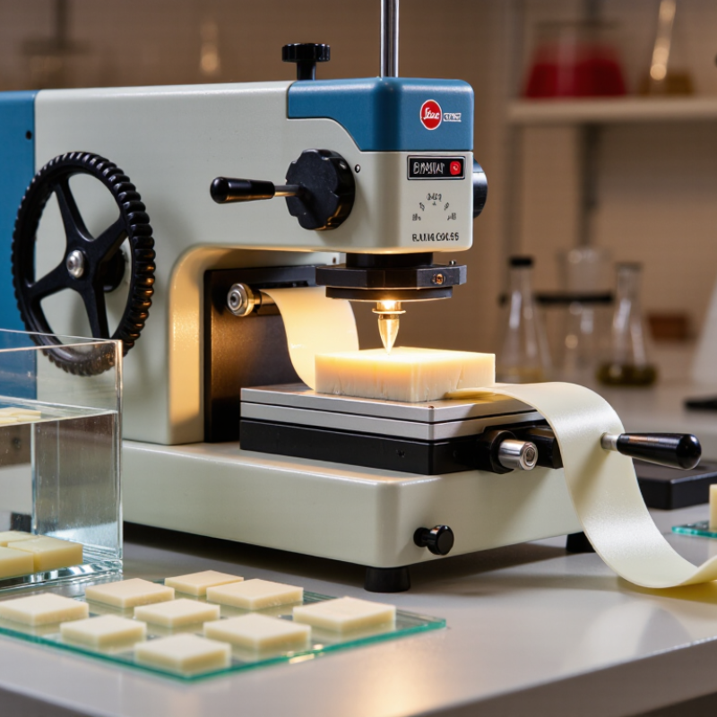
A rotary microtome is a laboratory instrument used to cut extremely thin tissue sections. It works with tissue blocks enclosed in paraffin wax. The operator advances the block toward a sharp disposable or reusable blade. Each rotation produces a controlled section, often between two and seven micrometres thick. These sections can be placed on glass slides for staining and microscopic examination. The core function is simple: create consistent slices without damaging cellular structure.
In routine histology, this instrument supports the study of biopsies, surgical specimens, and research samples. Good results depend on more than blade sharpness. The wax must be firm, the block face should be even, and the clearance angle needs careful adjustment. I have found that a slightly uneven block can cause ribbons to split or compress. It is not perfectly forgiving. Section quality should be checked under a microscope, not judged only by appearance.
Tips: Chill the wax block before trimming, but avoid excessive cooling that makes it brittle. Clean the blade holder after each session. Keep the cutting surface free from wax fragments. Use steady hand movement and consistent pressure. If sections wrinkle, review the block temperature, blade condition, and cutting speed before changing several settings at once. Small adjustments are safer.
A rotary microtome is used to cut thin, uniform sections of prepared specimens for microscopic examination. The chart shows representative section thicknesses commonly used for different laboratory applications.
How to read the chart: Routine paraffin sections are commonly cut at about 3–5 µm, while frozen sections are generally thicker. Resin-embedded specimens may be cut much thinner for higher-resolution microscopy. Actual settings vary with specimen type, embedding medium, and intended examination.
A manual rotary microtome is used to prepare thin tissue sections for microscopic examination. In routine histology, it cuts processed tissue blocks into consistent slices. These sections may be placed on glass slides for staining, diagnosis, or research.
The mechanism is precise but not automatic. A specimen block sits in a secure holder while the operator advances it toward a sharp disposable blade. Each handwheel rotation moves the block by a selected thickness, often between 1 and 10 micrometers. The operator controls cutting speed and pressure. A slow, even motion can produce a smooth ribbon. Uneven pressure may cause chatter, compression, or torn edges.
Small details matter. The paraffin should have the right hardness for the tissue. A cold block may cut cleanly, while an overly warm block can smear across the blade. Sections are floated on a warm water bath, then collected carefully to avoid folds. They may look perfect at first.
They are not always perfect.
Experienced technicians still check section thickness, tissue orientation, and blade condition under good lighting. A minor trimming mistake can remove an important tissue area. Regular cleaning also prevents wax buildup around the specimen clamp and feed mechanism. Safe handling is essential because the blade is extremely sharp. With careful setup and repeated inspection, this instrument helps create thin, representative sections that reveal cellular structure with dependable clarity.
| Data Dimension | Typical Value or Range | Practical Description |
|---|---|---|
| Primary Function | Thin-section preparation | A rotary microtome advances an embedded specimen toward a fixed blade so that a controlled slice is removed with each handwheel movement. |
| Main Laboratory Use | Histology and pathology | It is commonly used to prepare tissue sections for microscopic examination, including routine diagnostic staining and research analysis. |
| Typical Specimen Material | Paraffin-embedded tissue | Formalin-fixed, dehydrated, and paraffin-infiltrated tissue blocks are the most common specimens processed with a routine rotary microtome. |
| Routine Section Thickness | 2–5 µm | Sections in this range are widely used for standard hematoxylin and eosin staining because they provide a useful balance between cellular detail and structural integrity. |
| Broader Thickness Setting | Approximately 0.5–60 µm | Many general-purpose rotary microtomes offer a thickness-selection range covering very thin research sections through thicker sections for special applications; the exact range depends on the instrument configuration. |
| Section Thickness for Frozen Tissue | Commonly 5–20 µm | Frozen sections are normally produced with a cryostat rather than a standard room-temperature rotary microtome, because the specimen must remain frozen during cutting. |
| Section Thickness for Resin Blocks | Approximately 0.5–2 µm | Plastic or resin-embedded specimens can require specialized knives, clamps, and cutting conditions; ultrathin electron-microscopy sections generally require an ultramicrotome. |
| Cutting Principle | Controlled forward specimen feed | A feed mechanism moves the specimen a preset distance toward the blade. The selected feed determines the nominal thickness of each section. |
| Section Collection | Individual sections or ribbons | Paraffin sections may form a continuous ribbon when the block, blade, clearance angle, temperature, and cutting speed are properly controlled. |
| Common Blade Types | Disposable steel or reusable high-quality blades | Disposable blades are convenient for routine work, while reusable blades may be selected for particular block materials or specialized cutting requirements. |
| Mounting Surface | Microscope glass slides | Sections are transferred to clean slides, often using a water bath for paraffin sections, and are then dried or otherwise prepared for staining. |
| Typical Downstream Stains | Hematoxylin and eosin; special stains; immunohistochemistry | The thin sections provide access for routine morphological stains, connective-tissue stains, carbohydrate stains, microorganisms stains, and antibody-based labeling methods. |
| Specimen Size | Small tissue blocks | The specimen must be securely trimmed and clamped in a compatible block holder. Exact allowable dimensions depend on the holder and microtome design. |
| Key Preparation Step | Firm, uniform embedding | Complete infiltration and correct orientation help prevent compression, chatter, tearing, and uneven section thickness during cutting. |
| Important Operator Controls | Section thickness, cutting speed, blade angle, and specimen orientation | These controls influence section quality. A sharp blade, stable block, appropriate clearance angle, and steady handwheel motion are essential for consistent sections. |
| Typical Section Quality Indicators | Uniform thickness and intact morphology | Good sections should be sufficiently flat, continuous, and free from excessive compression, folds, knife lines, chatter, and block separation. |
| Primary Limitation | Requires a suitably hardened specimen | Soft, brittle, poorly infiltrated, or inadequately cooled specimens may not cut cleanly and may require modified processing or a different sectioning method. |
| Safety Considerations | Sharp-blade handling and handwheel control | The blade should be guarded whenever possible, the handwheel should be secured during blade changes, and waste should be discarded in an approved sharps container. |
| Resulting Microscopy Benefit | Improved light transmission and cellular resolution | Thin sections allow transmitted light to pass through the tissue, making cellular organization, nuclei, connective tissue, and pathological changes easier to examine. |
A rotary microtome is used to cut thin tissue sections from paraffin-embedded blocks. In routine histology, technicians commonly produce sections around 3–5 micrometres thick. These ribbons are placed on glass slides, stained, and examined under a microscope. The instrument supports cancer diagnosis, biopsy review, surgical pathology, and teaching collections.
The need is substantial. IARC’s GLOBOCAN 2022 report estimated 20 million new cancer cases and 9.7 million cancer deaths worldwide. Accurate tissue preparation therefore matters. Consistent sections help pathologists assess cell structure, tumour margins, inflammation, and tissue damage.
Researchers also use serial sections for immunohistochemistry, tissue morphology, biomarker studies, and some molecular workflows. Small differences matter.
In practical laboratory work, cutting quality depends on blade condition, block temperature, tissue hardness, and operator technique. A thin section is not automatically a good section. Compression, chatter, folds, or uneven thickness can weaken interpretation and waste valuable samples. The instrument is useful for routine paraffin work, but it requires disciplined handling and regular quality checks. ASCO and CAP guidance continues to emphasize reliable specimen processing and standardized pathology workflows, although local procedures may differ. That variation deserves attention. A well-maintained microtome can support repeatable results, yet human judgment remains central at every slide.
What Is the Leica RM2235 Microtome Used For?
Main Components Supporting Precise Sectioning
A rotary microtome is used to cut thin tissue sections from paraffin blocks. These sections support routine histology, cancer diagnosis, and research microscopy. The 2022 WHO/IARC Global Cancer Observatory estimated 20 million new cancer cases worldwide. That volume highlights the need for consistent, traceable slide preparation.
The specimen clamp holds the embedded block firmly during cutting. A handwheel moves the block through a controlled vertical stroke. The feed mechanism advances the specimen by a selected thickness, often around 2–5 micrometres for routine paraffin work. A rigid blade holder helps maintain a stable cutting angle. The anti-roll plate can guide the ribbon across the blade, reducing folds and compression. Small errors matter.
The coarse-advance control brings the block toward the blade quickly, while fine feed supports the final sectioning stage. A lock on the handwheel improves safety during blade changes and cleaning. The waste tray also matters because loose ribbons can contaminate nearby surfaces. ISO 15189 emphasizes competence, equipment control, and reliable laboratory processes, so daily checks should include clamp security, feed movement, and blade condition. In practice, even experienced operators may adjust pressure, clearance, or paraffin temperature repeatedly. That is the weak point. Precise components cannot fully correct a poorly processed block or rushed handling. Records of section thickness, maintenance, and unusual resistance make later review more credible.
A rotary microtome is used to cut thin tissue sections for microscopic examination. It supports diagnostic pathology, research, and teaching laboratories. Its value depends on controlled operation, not simply sharp blades. The operator must secure the specimen, select suitable clearance, and advance the block gradually. Uneven feeding can create chatter, compression, or incomplete sections.
Safe use begins with a documented risk assessment. The World Health Organization’s Laboratory Biosafety Manual, fourth edition, recommends assessing hazards before work and reviewing controls when procedures change. Keep fingers behind the blade holder, use the handwheel lock during adjustments, and remove disposable blades with forceps or a dedicated tool. The U.S. Bureau of Labor Statistics recorded about 2.6 million nonfatal workplace injuries in private industry during 2023. That figure covers many sectors, but it reinforces a practical point: routine tasks still require disciplined controls. The workflow is not perfect. Rushing during trimming remains a realistic weakness.
Tips: Inspect the clamp before sectioning. Confirm the block is frozen, fixed, or embedded correctly. Use a clean brush, never a fingertip, to move sections. Record unusual resistance, vibration, or repeated tearing. Stop when the blade edge is damaged. Clean exposed surfaces after work, following your laboratory’s approved procedure. Training should include supervised practice, emergency response, and periodic competency checks. A second review can reveal habits the operator no longer notices.